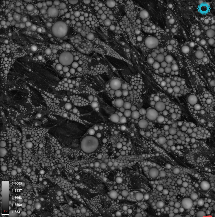
Holotomography (HT) is the most versatile label-free live cell analysis platform providing unprecedented precision of three-dimensional subcellular information.
The now on-demand webinar introduces Tomocube’s HT-X1, the first ever holotomography technique to use a low-coherent light source with multiple beam patterns to obtain quantitative 3D Refractive Index (RI) information thereby minimizing interference noise, and eliminating the need for a calibration step for image acquisition.
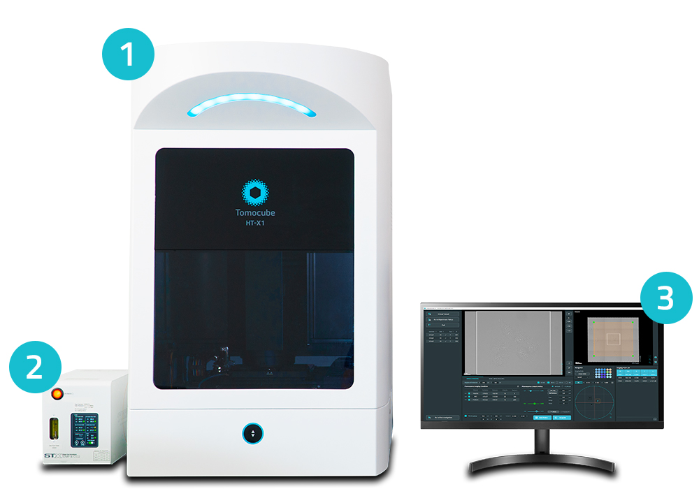
The motorized stage allows for tiling and multi-point analysis within each well, in addition to moving between wells. A built-in incubation system completes the live-cell imaging setup. In addition to quantitative RI information, users can correlate holotomography images with 4 channel of correlative 3D fluorescence. The applications are similar to other holotomography imaging systems, although the HT-X1 is an especially powerful tool for:
Confluent & sensitive live cells: primary cells including stem cell and neuronal cells,
Monitoring of multiple samples
Observing thicker samples
Nanomaterial delivery: better resolution without speckle noise
We at Nexus would like to wish everyone a happy new year, and announce that we will be attending the Advanced Imaging Methods Conference at UC Berkeley this January 24th – 26th. If you haven’t registered yet, the opportunity is still available here. We are excited to showcase Tomocube’s new automated, label-free imaging system, the HT-X1.
For those of you who would like to learn more about the HT-X1, we will be hosting a pre-conference webinar to highlight the event and the system this Friday the 13th at 11am PST. Register here – even if you can’t make the conference it will still be a great opportunity to learn more! The MIC at Berkeley has also given us a dedicated space to house the system for the week surrounding the conference. Live demonstrations will be held for any interested users from January 18th – February 3rd. Don’t miss your chance to test your samples and book a personal visit. See below!
Maximum-Intensity Projection Video of a Label-Free Holotomographic Image
The HT-X1 is the latest of Tomocube’s label-free microscopes. The system adapts the ODT technique to achieve compatibility with standard multi-well plates, while still retaining the many advantages of label-free holotomography. With a large capture field, multi-point and stitching capabilities, the HT-X1 scales the amount of data acquisition by multiple orders of magnitude. 3D fluorescence capabilities and a fully integrated incubation system provide true multi-dimensional imaging across time, space and modality
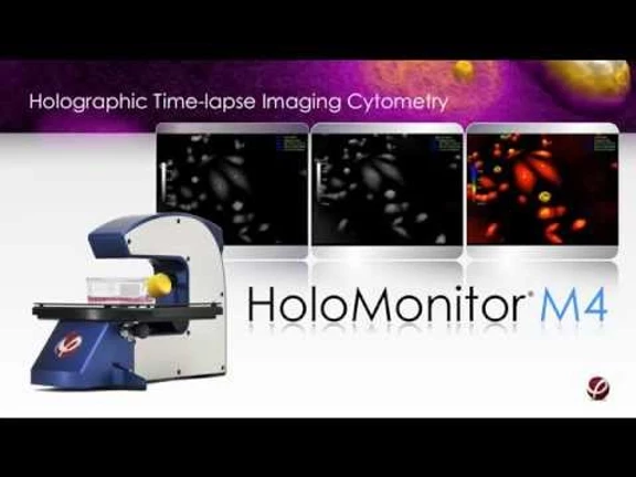
PHI’s (Phase Holographic Imaging) non-invasive HoloMonitor® M4 lets you continuously image, monitor and, analyze single cells as well as cell populations directly inside the incubator without any labels or stains. It is a compact microscope that allows continuous digital holography imaging inside an incubator for tracking cells in real-time.
The Holomonitor® App Suite Software is an integrated solution with cell biological different application modules including:
Guided End-point Assays to quickly assess cell count and cell culture quality
Cell counting
Cell quality control
Guided Kinetic Assays for kinetic results that are publication-ready
Kinetic Cell Proliferation
Kinetic Cell Motility
Kinetic Dose Response
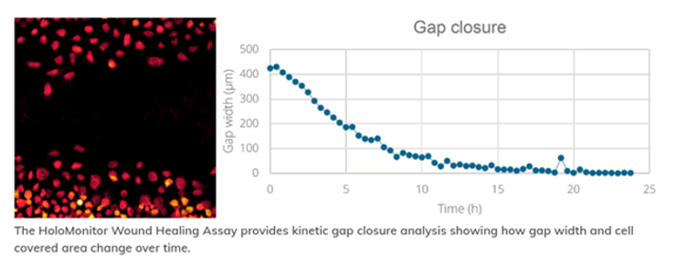
Wound Healing
In-dept Analysis Assays for single cell tracking for the analysis of individual cell and cell populations
Single cell tracking
Cell morphology
Automatic single cell tracking saves time
The Holomonitor® AppSuite software tracks all cells with great precision, including cell division detection and assigning daughter cells to their cell family. This gives you insights into the entire family tree of a cell without any cellular markers.
The digital holography enables automatic collection of data on an individual cell within the image, giving insights in tracking, cell behavioral, or morphological changes.
Analyze both cell movement and morphological changes
With Holomonitor®,you can follow cells over time inside the incubator. With a single sample, multiple behavioral and morphological changes in the cell culture can be studied. In a single experiment, you can examine cell movement plots over time as well as study the changes in cell morphology features within a single celland/ora cell population.
Improved data export allows deeper insights
With the App Suite software, you can export the single cell tracking analysis into a spreadsheet for further analysis. This data allows the study of morphological features, as well as cell cycle length etc.
Benefits of HoloMonitor® for Live Cell Imaging
Cut down on analysis time, costs and valuable cells
Expensive consumables are not needed
Operation is label-free
No phototoxicity
Cells can be used for other experiments after imaging
Export data to a spreadsheet with a single click
Faster tracking adjustments
Optimized non-biased results
Automated analysis keeps user bias to a minimum
Continuous cell monitoring
Real-time results
Direct, quantitative measurements
Whether you need live cell imaging for single cells or cell populations, read more about the PHI Holomonitor® at Nexus Scientific or call (857) 217-0936.The PHI Holomonitor® allows continuous imaging, monitoring and quantitative analysis of both single cells and populations of cells directly inside the incubator without any labels or stains.
Sports have been played for ages. However, competitive sports require enhanced athletic performance to improve the odds of winning. Athletes and coaches are always looking to improve sports performance. Enhancing performance requires a thorough understanding of many internal or external factors.
Most athletes are aware of internal factors, like genetics and muscle fiber type, and external factors, like the environment. But research shows us there is more to performance. One such factor is ‘Oxidative Stress’.
Why is oxidative stress harmful?
Oxidative stress refers to the difference of free radicals and antioxidants in the body. Free radicals are molecules that easily react with other molecules, causing oxidation reactions. These are usually balanced by antioxidants in the body. But when there’s the free radical activity far exceeds the antioxidant activity, you experience oxidative stress. As a result, the free radicals start affecting lipids, DNA, and proteins, causing premature aging or diseases, like heart disease, diabetes, inflammation and more.
Sports and exercise increase the production of free radicals. Exercise leads to an increased demand for oxygen, especially in the muscles, altering blood flow to the various organs. Exercise also causes muscle injury, leading to entry of phagocytes at the site. All these physiological changes add to the production of free radicals, causing oxidative damage to biomolecules.
An interesting fact to know is a moderate, short-term increase in free radicals is good for the body as it aids physical adaptation. This is possible with a good physical activity program. But excessive oxidative stress could impact an athlete’s performance in several ways.
Muscle damage
Muscle injuries are common while training. But did you know that damaged muscle fibers cause infiltration of phagocytes that break down the damaged tissue and further attract more white blood cells to the site.
This can ideally cause regeneration of muscle fibers. However, in case of excessive activity, it may lead to chronic inflammation, improper healing, and, in some cases, scar-tissue formation.
Motor skills
Retired professional athletes, especially those who played contact sports, have been seen to be at a higher risk of neurological diseases, like Alzheimer’s and ALS.
Atherosclerosis
Cholesterol is required in the production of several biomolecules, such as hormones, and coenzymes. It becomes harmful when free radicals oxidize it. In that case, it gets embedded in walls of the blood vessels, preventing normal blood flow. A combination of this and oxidative stress can lead to atherosclerosis.
Can Oxidative stress be prevented?
Oxidative stress is necessary to some extent, so it can’t be completely eliminated from sports. As an athlete, the next best thing is oxidative stress tracking for recovery throughout a training cycle to make sure you are creating positive adaptations without causing cell damage.
Fortunately, the O2Score device, available at Nexus Scientific, makes it possible to scientifically quantify the body’s response to training. Athletes and coaches can make sure the training loads and recovery methods are optimal, and performance can be maximized, without causing excessive oxidative stress and the risk of injury or fatigue.
What is oxidative stress and why is it important for athletes?
A balance between free radicals and antioxidants for optimal physiological functioning of the body. If free radical production exceeds the amount or the speed at which the body can regulate them, you experience oxidative stress. Simply put, free radicals impact lipids, proteins, and DNA, leading to several ailments in the long run.
Free radicals – the rogue molecules
Free radicals are produced in large quantities in muscles during exercise. These moleculesmay be created when ATP (energy) is produced through mitochondria in our cells. Another source of free radicals is the environment – pollution, alcohol, tobacco smoke, heavy metals, transition metals, industrial solvents, pesticides, paracetamol, and radiation.
What happens during exercise?
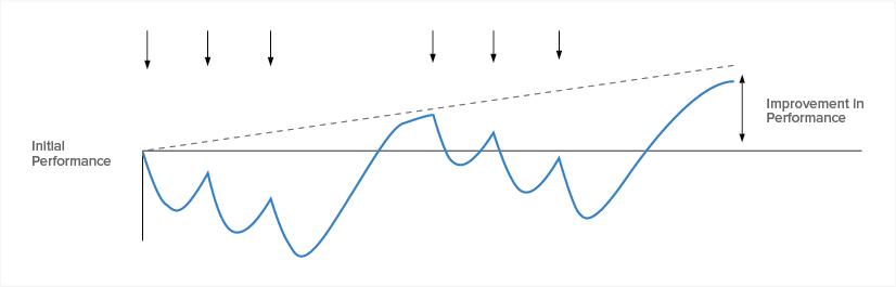
During exercise, the body creates free radicals, followed by antioxidants to combat these free radicals. However, high intensity or exhaustive exercise may skew this balance, not allowing your cells to recover in time. As a result, you may experience fatigue, cognitive impairment or brain fog, muscle and joint pain when you exercise again the next day.
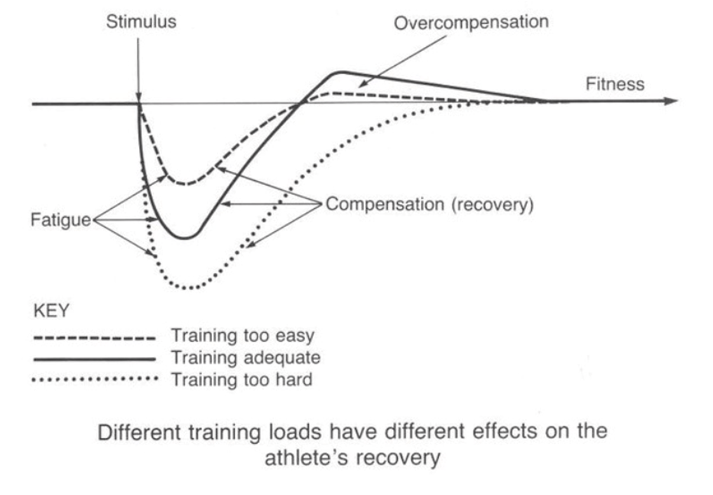
However, this is not a bad thing! When you train harder before the body adapts, overcompensation occurs to prepare the body for the next time it’s exposed to the same stimulus. The problem really lies in continuous overtraining, without giving the body enough time to recover and adapt. It would result in a buildup of excess free radicals that bind to lipids, proteins and DNA creating fatigue, joint and muscles pain, injury or cognitive impairment (brain fog), and eventually cell death (apoptosis).
In contrast, if the body is not exposed to an overload or you go through a long recovery period, your body will not undergo much overcompensation, will not adapt and your training will plateau because you will not have created enough oxidative stress!
Why does oxidative stress matter?
Oxidative stress is a scientific measure correlated to how well recovered you are.
Understanding your body and knowing its response to different training stimuli, is key to reducing the risk of injury and obtaining a steady progression in fitness. You can supplement your recovery by increasing antioxidant power through your diet. Methods of increasing blood flow such as stretching, foam rolling, cold contrasting, compression and elevation, are all good ways to help deliver nutrients to your muscles. Getting good quality sleep will also allow for those adaptations to take place. Keep a training log with an RPE (rate of perceived effort), so you can adjust your training load depending on how you felt.
Athletes and their coaches can scientifically track their oxidative stress throughout the training cycle to make sure they are creating positive adaptations without causing cell damage. Now you can measure your antioxidant power with the O2Score device and its interactive mobile app.
In reality, you may workout more than one day in a row before you take an easy day. What is important is to make sure you are recovering sufficiently between ‘key’ workouts and supplying the body with the right amount of stimulus during these workouts, creating a progressive overload. Too little stimulus will lead to no adaptations and too much will lead to fatigue and injury. If you are looking for oxidative stress tracking for recovery, check out theO2score device at Nexus Scientific or contact info@nexusscientific.com.The O2Score is a robust pointer of the antioxidant power, made after extensive R&D carried out at EPFL (Switzerland), validated for several years by professional athletes and coaches in various disciplines.
Phase Holographic Imaging’s Holomonitor is a compact label-free live cell imaging cytometer allowing non-invasive quantification and visualization without compromising sample health. The system operates in an automated format and is designed to fit inside standard incubators. The results are non-invasive real 3D images and a range of quantitative population-level data of cell cultures, all the way to single-cell analysis. You can follow PHI’s webinars and blog posts for the most up-to-date information on the Holomonitor system and its user applications.
Follow the link below for one of the most recent blog posts published by PHI:
HoloMonitor User Spotlight
Hear directly from current users within multiple research areas and applications about how quantitative live cell imaging data is useful to them.
Monica Hellesvik, a Ph.D. candidate at the University of Bergen in Norway, shares how she uses HoloMonitor as her main method to study cancer cell behavior.
CPD-017 by Kataoka Corporation to Streamline iPSC Processing for the US Market
Los Angeles, California, May 25th, 2022 – Kataoka Corporation (Kyoto, Japan) and Kataoka-SS America Corporation (Carson, CA) have developed the CPD-017, a cell processing platform that uses cutting edge laser and AI technology to purify adherent iPSC cultures.
The CPD-017 uses a platform of integrated technologies to process iPSC samples; built-in phase contrast and fluorescence microscopes to image samples, a tailored, machine-learning AI model to process the images and identify spontaneously differentiated cells, and laser treatment to target discrepant cells with minimal stress to the surrounding environment.
The CPD-017 is now available for purchase – Kataoka Corporation is pleased to announce its first US-based customer, the Cedars-Sinai Medical Center Biomanufacturing Center, a division of the Board of Governors Regenerative Medicine Institute.
More information about Kataoka Corporation and the CPD-017 can be found at https://www.kataoka-ss.co.jp/en/. Requests for further product and sales-related information can be submitted to salesusa@kataoka-ss.co.jp, or through the inquiries form on the website.
About Kataoka Corporation
Kataoka Corporation, headquartered in Kyoto, Japan, with U.S. offices in Carson, CA and Reno, NV, is a leader in precision laser technology, and has applications in various niches including charge-discharge battery inspection systems, and Life Sciences in addition to laser drilling and welding machines.
The webinar on Phototoxicity will focus on presenting a recent study realized in collaboration with Ecole Polytechnique Féderale de Lausanne (EPFL). The research compares standard epifluorescence microscopy with the Nanolive label-free tomographic phase imaging technique and discusses the perturbations induced by phototoxicity on living samples during experimentation.
In particular, the following topics will be covered:
The challenge of observing live cells unperturbed
The effect of light exposure on living samples
Nanolive imaging to observe unperturbed organelle movements
Overcoming Phototoxicity - The Nanolive Imaging Advantage
In vitro live cell imaging has become a promising tool to study dynamic cell behaviors, such as division and differentiation, cell death, cell-cell interaction (e.g. immune cells), or cell response to treatments over extended periods of time. However, few technologies have been developed with phototoxicity in mind. Today, most live imaging techniques rely on either high illumination regimes or fluorescent labelling, both inducing phototoxicity and compromising the ability to keep cells unperturbed and alive over time. Since our knowledge of biology is driven by observation, it is key to minimize the perturbations induced by the imaging technique.
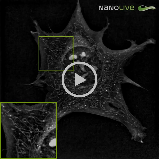
Surprisingly, very few studies have highlighted or discussed the artifacts arising from phototoxicity during live cell imaging. This page focuses on the Master Project of Hugo Marc Moreno, which compares standard epifluorescence microscopy with Nanolive imaging (also known as holo-tomographic phase microscopy). Herein, Moreno et al discuss the perturbations induced by phototoxicity on living samples during experimentation.
Nanolive Imaging gets rid of invasive fluorescent markers, while using a low energy exposure light source. Nanolive’s label-free imaging modality makes it well suited to observe living samples, when compared to epifluorescence imaging. Moreno’s et al results raised the question of how much of our knowledge, particularly about mitochondria, may have been biased by artifacts induced by fluorescence imaging.
Nanolive microscopy alone did not induce any detectable effect on mouse pre-adipocytes after one-hour acquisition at a frequency of 1 image every 6 seconds. The overall spreading and motility of the cell remained apparently uncompromised: cell shape did not drastically change nor shrink over the imaging period, the ability of filopodia to spread and adhere looked unaffected and main adhesion sites were not impaired. Shape and density of nucleoli (bright granules in the nucleus) did not provide any sign of perturbation neither. Moreover, mitochondrial network did not show any change in phenotype: no extended fusion nor fission was observed, the overall dynamic of the mitochondria remained constant during the full hour of acquisition, and the shape of mitochondria was conserved, no swelling of mitochondria was seen.
Imaging regimen: 1-hour acquisition at a frequency of 1 image every 6 seconds
VS
Cells are affected very rapidly:
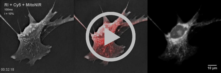
Appearance of round-shaped or swollen mitochondria was observed with Nanolive technology.
Loss of mitochondrial specific fluorescent signal and high background or cytosolic fluorescent signal could be observed on the fluorescent image.
Mitochondrial dynamics slowed down and reached an almost complete stop after 38 minutes of exposure.
Very slow cell shrinkage was observed until timepoint 00:58:00, at which a consequent proportion of the cell lost adherence.
MitoNIR signal was concentrated in the nuclear periphery at later timepoints.
If you are interested in learning more, look into Moreno’s et al. Master thesis here.
The Latest in Nanolive Imaging: CX-A
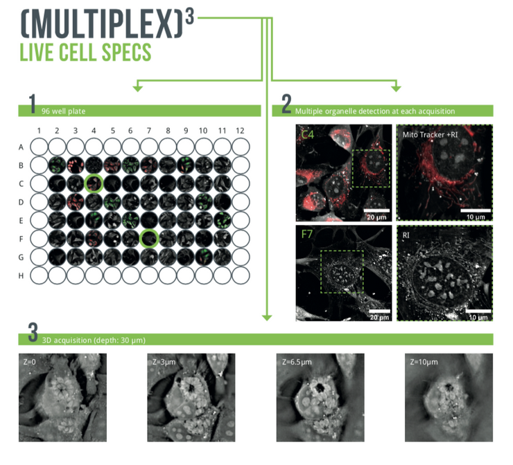
The CX-A redefines the limits of live cell imaging in 96 well plates for continuous organelle monitoring in cell populations.
Play Video
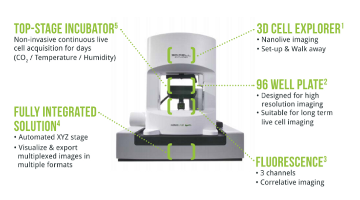
Complete Solution for 3D Live Cell Exploration
The CX-A is designed to work with 96 well plates to multiply and parallelize experimental conditions, hence, bringing undoubtable significance to each experiment and delivering solid biological insights to researchers (1).
Furthermore, the system is equipped with multiple imaging modalities to correlate and compare physical and chemical information at each time-point (2).
Finally, 3D data sets of every single image at every single time-point are automatically acquired in real-time (3).
A fully integrated solution adapted to the most advanced professional needs. Click here for a full overview of applications.
The Nexus Team wishes you a happy and healthy Fall. As a Life Science Service business, Nexus continues to provide distribution of innovative products in the United States. Here are the latest updates from us, including:
New Socially Distanced Demo's
Part 2 of Live Cell Webinar Series: STEM Cells
The W8 on our New Product Radar for Measuring Organoids
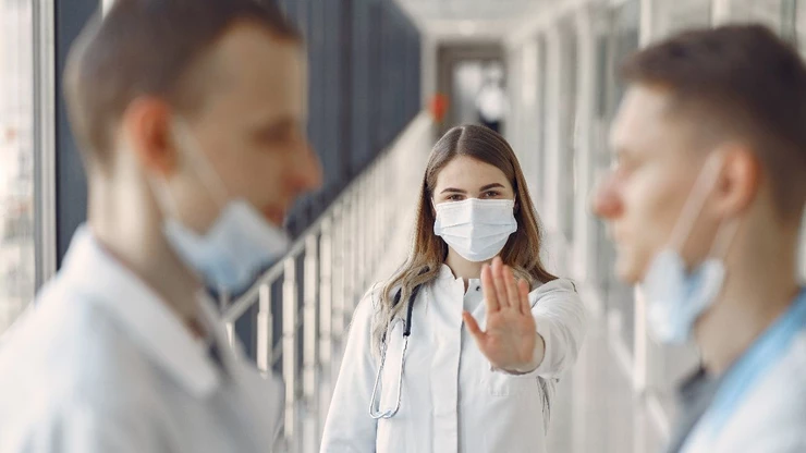
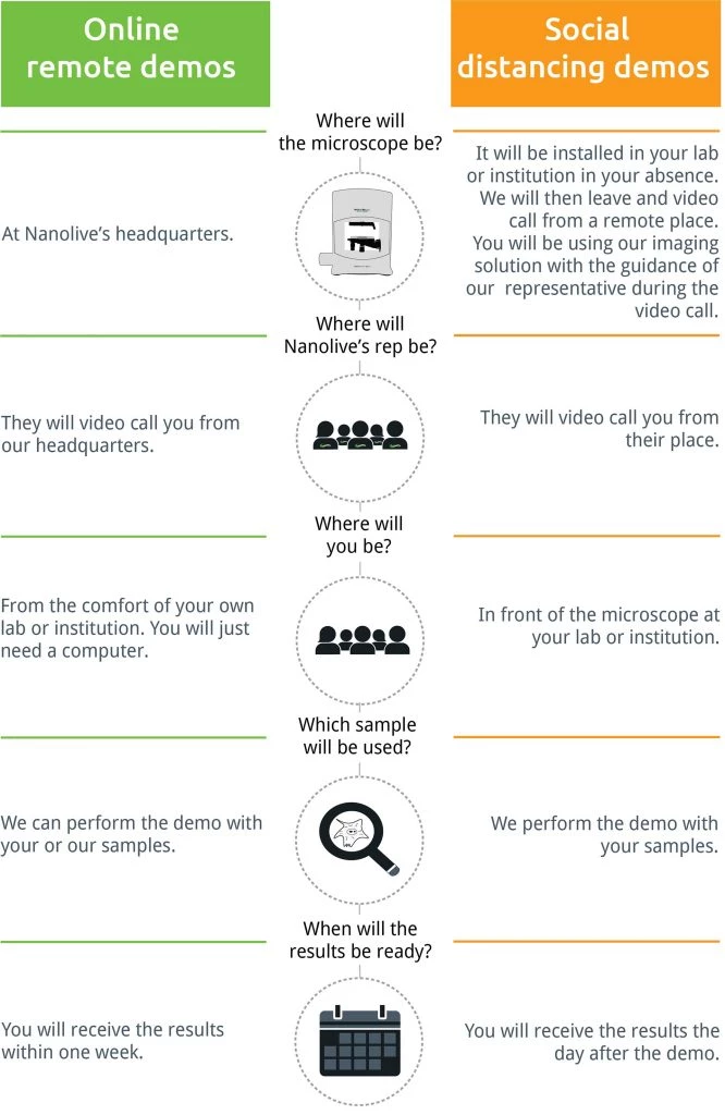
New! Socially Distanced Demonstrations
During these fast changing times, Nexus is striving to provide the best support to current and future clients. From virtual to onsite (small group) demonstrations, we offer a wide variety of support for our customers. If you are interested in live demonstrations from the Nanolive or PHI AB product lines, please complete the short survey.
Here's How it Works:
A New Approach to Measuring Organoids and Spheroids
The W8: A Physical Cytometer for 3D Samples
Nexus is asking all organoid and spheroid experts for their take on a new system in development, the W8 by CellDynamics. The system gathers precise information about size, weight, and mass density on 3D cultures from single cell suspensions to spheroids. Please contact us with any feedback or questions.
Complete Solution for 3D Live Cell Exploration
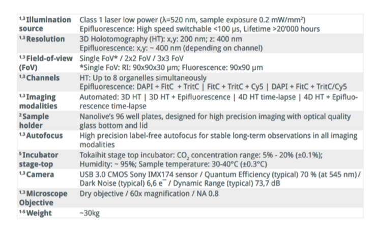
Nanolive Imaging combines automatic 3D refractive index analysis with a fully integrated 3-channel epifluorescence module to image live cells as long as you want, providing label-free imaging for new biological insights. Nanolive’s latest product, the CX-A, automates this imaging across a 96-well plate layout for statistically relevant, high-resolution data.
Label-Free Imaging Without Leaving the Incubator
Phase Holographic Imaging's HoloMonitor M4
Nexus Scientific has recently partnered with Phase Holographic Imaging (PHI), developers of the Holomonitor. Designed to fit inside standard incubators, the Holomonitor is able to non-invasively explore of the kinetics of cellular responses on a population level. The label-free imaging system is based on the principle of quantitative phase imaging, enabling non-invasive visualization and quantification of living cells without compromising cell integrity.
We at Nexus are currently offering virtual presentations and webinars for both systems, as well as organizing socially distanced demos for the future. Contact us for more information.
The Nexus Team wishes you a healthy and productive start to the Fall season. As a Life Science Service business, Nexus continues to provide distribution of innovative products in the United States.
Phase Holographic Imaging's HoloMonitor M4
Nexus Scientific has recently partnered with Phase Holographic Imaging (PHI), developers of the Holomonitor. Designed to fit inside standard incubators, the Holomonitor is able to non-invasively explore of the kinetics of cellular responses on a population level. The label-free imaging system is based on the principle of quantitative phase imaging, enabling non-invasive visualization and quantification of live cells without compromising cell integrity.
Join us for a Webinar on Truly Controlled Cell Experiments for Cancer Research
Phase Holographic Imaging (PHI), in collaboration with Scientific Bioprocessing and BioSpherix, is hosting the first of a three-part webinar series dedicated to enhancing the translatability of in-vitro findings. All three technologies are designed to limit artifacts, increase reproducibility, and provide quality control to ensure that experiments are conducted in an environment that it as close as possible to the true physiology. The first webinar is aimed at cancer research and will be held on October 14th at 11am EST. Click here to register and learn more.
The Speakers
Phase Holographic Imaging (PHI)
Provides a non-invasive tool that lets you continuously image and quantitatively analyze both single and populations of cells directly inside your incubator without any labels or stains.
Scientific Bioprocessing
SBI’s real-time pH and dissolved oxygen sensors monitor pericellular conditions with tiny sensors as small as 3 mm in diameter. Sensor feedback can be used to control agitation so that cultures can reach desired dissolved oxygen levels with the ID·Rocker instrument.
BioSpherix, Ltd.
Designs and builds Cytocentric equipment for academic research, pharmaceutical, and biotechnology laboratories around the world. Our equipment uniquely supports the needs of cells for constant physiologic conditions, increasing reproducibility for cell-based sciences and therapies.
Automated Gap Closure Analysis for Wound Healing
PHI provides a number of different automated analyses for applications such as wound healing (below), cell viability, cell proliferation, cell tracking, drug response, and more! We at Nexus are currently offering virtual presentations and webinars of the system, as well as organizing demos for the future. Contact us for more
Thank You
Your form has been successfully submitted.
Get A Quote
Kataoka Cell Processor
PHI Downloadable Website Brochures
Our Spring Sale Has Started
You can see how this popup was set up in our step-by-step guide: https://wppopupmaker.com/guides/auto-opening-announcement-popups/