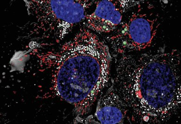
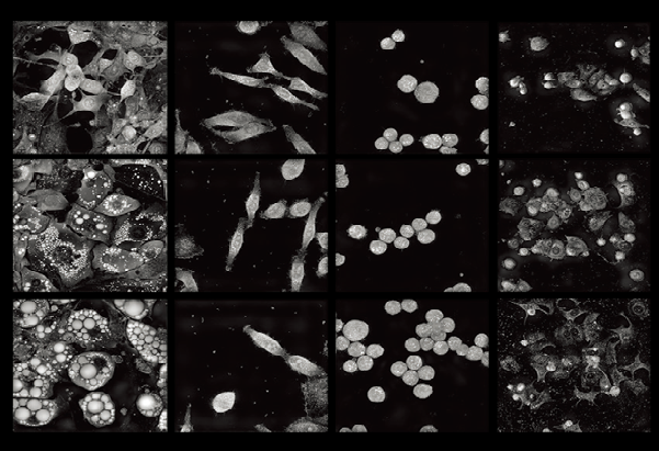
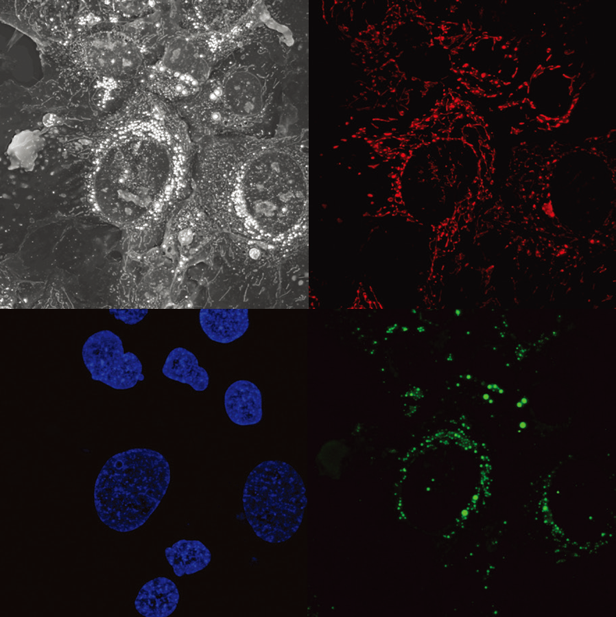
The challenge of live-cell imaging is being able to visualize your samples while causing as little perturbation as possible. Fluorescence techniques generally have to balance the rate of acquisition against the total duration of the experiment in order to preserve sample health. Holotomography is a non-invasive, label-free technique that allows visualization at high resolution, while maintaining the cell’s natural state. It does this by allowing very low levels of light to pass through the sample at multiple angles and measuring the refractive index (RI) of various subcellular structures.
By taking information from multiple angles, the Tomocube systems take all of their measurements in 3D. This allows quantification of values such as volume, surface area, and dry mass of the cell and intracellular structures.
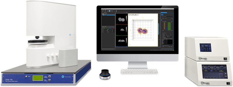
HT-X
The HT-X is Tomocube’s first high-throughput Holotomographic microscope. It combines all of the previous advantages of Tomocube’s system and adapts them into a multi-well plate format. Like the former models, it is capable of 3D label-free imaging at high resolution and 3D fluorescence imaging. The motorized stage allows for tiling and multi-point analysis within each well, in addition to moving between wells. A built-in incubation system completes the live-cell imaging setup.
The HT-2H is the second iteration of Tomocube’s Holotomographic microscopes. Like its predecesser, the system is capable of label-free 3D imaging at a high resolution. It is then paired with an external fluorescence module capable of taking 3D fluorescence z-stacks. This allows you to overlay your label-free data with traditional fluorescence information. The motorized system is capable of tile-stitching and multi-point analysis, as well as compatible with a stage-top incubator to control environmental conditions.
The HT-1H is Tomocube’s first generation of Holotomographic microscopes. It is capable of high-resolution, label-free, 3D imaging at 2.5fps. A motorized stage allows for tile-stitching and multi-point analysis, and a stage-top incubation chamber completes the live-cell imaging setup.