Key Partner PHI Begins Trading on the OTCQB Venture Market in the U.S.
A key partner for Altium, Phase Holographic Imaging (PHI), has officially announced that it’s trading on the OTCQB Venture Market under the symbol “PHIXF” as of August 15th. This listing marks a significant achievement in PHI’s mission to expand its presence and engage more effectively with U.S. investors.
For our customers and community, this development represents a strengthened commitment to the North American market. With PHI’s listing on OTCQB, U.S. and Canadian customers can expect increased accessibility and transparency, making it easier to stay informed about the company’s growth and innovations. At Altium, we are fully prepared to meet the anticipated rise in demand for PHI’s cutting-edge holographic imaging products.
Our readiness to supply PHI’s products means that customers can continue to rely on Altium as their trusted partner in the rapidly advancing field of holographic imaging. We are committed to ensuring that our research community has access to the latest technologies and support they need to succeed.
This step forward underscores PHI’s dedication to expanding its reach and engagement with the North American user-base, paving the way for future growth and collaboration.
For more information on the PHI M4 Holomonitor, download the Holomonitor brochure or contact us at info.us@altium.net.
We are thrilled to share a significant development that marks an exciting chapter in our journey. We are excited to announce that Nexus Scientific is now a part of the Altium family. This strategic move brings a wealth of new opportunities and avenues for us to better serve you. As a result of this acquisition, our company’s name will transition to Altium International Inc.
For the past several years, Nexus Scientific has been your go-to partner for cutting edge life science research equipment. Our commitment to bettering our community has been the driving force behind our offerings, catering to your unique needs in the ever-evolving landscape of laboratory research.
With a product range spanning advanced imaging platforms to cutting-edge cell research platforms, we have been at the forefront of facilitating breakthroughs in cellular research and beyond.
Starting from August 2023, all official communications, invoices, and interactions will carry our new name, Altium International Inc. Despite this change, our core values, the expertise of our team, and our unwavering dedication to supporting your pursuits remain unchanged.
It’s important to note that this transition will not impact our team, management, or our operations. We will continue to operate from our existing locations, ensuring that you receive the same exceptional service you’ve come to expect from us.
If you have any questions or need further information about this transition, please don’t hesitate to reach out to selen.oztunaoglu@altium.net or (857) 264 6884. Our team is here to provide the answers you seek and the support you deserve.
Your trust and partnership have been pivotal to our growth, and we want to thank you for your continued support. We look forward to serving you even better as Altium International Inc., and we invite you to embark on this exciting journey with us.
Thank you for being an integral part of Nexus Scientific’s story, and we can’t wait to continue this journey together as Altium.
Assistant Professor, University of Michigan-Dearborn, USA
An interview with Dr. Besa Xhabija, Assistant Professor, University of Michigan-Dearborn, USA, as she discusses her use of the HoloMonitor system and AI tools in cancer research. She describes how non-invasive time-lapse imaging aids her breast cancer cell studies, allowing her to observe cellular behaviors and morphological changes affected by natural compounds and environmental toxins. The interview also delves into her recent publication, Kaniski et al. (2024).
Dr. Xhabija elaborates on how she incorporates the system into her workflow, enhancing both efficiency and data quality. She highlights the advantages of utilizing AI to maximize the extensive data generated by HoloMonitor. The conversation covers both technical aspects and broader implications, exploring how single-cell analysis and AI tools could significantly impact cancer research and regenerative medicine.
Altium Partners with Genmark to bring their geneMAP PCR Diagnostic Kits to the US
We are thrilled to announce our first product family offering in the clinical and diagnostic space, reflecting our dedication to expanding our portfolio to offer a wide range of products to our Life Science community. As of April 2024, Altium has become the US supplier for Genmark’s comprehensive portfolio of GeneMap PCR Diagnostic Kits. We look forward to working closely with diagnostic labs across the US to provide high-quality products at competitive prices.
Genmark Saglik is renowned for advancing molecular diagnostics and providing high-quality solutions that guide critical healthcare decisions. With over 60 products Genmark’s expertise spans oncology, genetics, and microbiology. This partnership aligns with our commitment to delivering top-tier diagnostic solutions to healthcare professionals across the United States.
For more information on our product offerings, download geneMAP Brochrue or contact us at info.us@altium.net.
Altium Expands Into in vivo Imaging with Vieworks Partnership
We are proud to announce that as of January 2024, Altium has partnered with Vieworks to represent their Bio Imaging product portfolio in the US and Canada. Vieworks is a leader in advanced imaging technologies, offering high-performance solutions such as their VISQUE® InVivo Systems for preclinical imaging and LUCEON digital pathology slide scanners. This collaboration marks another milestone in our commitment to providing state-of-the-art tools to the research sector in the pre-clinical space.
This collaboration introduces the VISQUE® InVivo Systems, which are advanced preclinical in vivo imaging solutions. The VISQUE® InVivo Series offers high-resolution fluorescent and bioluminescent imaging with features like -90°C cooling for enhanced sensitivity, time-lapse imaging, and intelligent analysis software, making it ideal for applications such as drug biodistribution, pharmacokinetics, and tumor tracking.
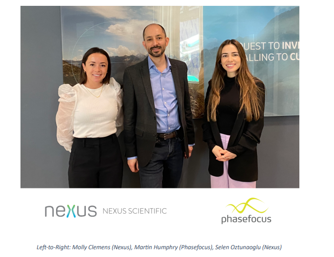
Today, Phasefocus and Nexus Scientific announced a partnership to accelerate the adoption of Livecyte, a high content live cell analyser, in the US market. Live cell imaging is essential to answering some of the most pressing questions in the life sciences, but conventional methods often involve labeling cells, a potentially harmful process that can alter research outcomes. Livecyte’s integration of patented label-free Quantitative Phase Imaging technology with state-of-the-art automatic cell tracking algorithms enables users to automatically characterize growth, morphology and motility of large populations of cells in a 96-well plate assay format.
Nexus brings extensive experience in the cell analysis sector to the partnership, with a portfolio of cutting-edge live cell imaging and analysis tools. With the addition of Livecyte, Nexus can offer researchers a unique depth of live cell analysis capabilities in application areas such as drug discovery, regenerative medicine and cell-based assays.
Phasefocus CEO, Martin Humphry commented: “We are very excited to be embarking on a new adventure with the fantastic team at Nexus Scientific. Their extensive sector knowledge and talented team are a great complement to Phasefocus. The partnership will help us grow our US customer base and bring the unique capabilities of Livecyte to a new audience.”
“We are proud to be adding the ground-breaking Livecyte to our growing live cell imaging and analysis portfolio” added Selen Oztunaoglu, Managing Director at Nexus Scientific. “With Livecyte’s unique and powerful capabilities, we get to address a wider range of customers’ pain points and they’ll will be able to enjoy new depths of insights and push the boundaries of knowledge even further.”
About Nexus Scientific
Nexus Scientific is a Life Science Equipment Supplier headquartered in Boston, MA. We provide International Trade, US Distribution of Equipment as well as pre-sale to post-sale support. Our customers range from Academic Research groups to Hospitals, Biotech and Pharmaceutical companies.
Working with Innovative brands and disruptive technologies is at the core of Nexus’s partnerships. Its mission is to enable scientists to further their research by ensuring ease of access to leading edge laboratory and research equipment. Nexus Scientific provides an opportunity for innovative life science brands to grow their global presence.
Phasefocus is changing what is possible in live cell assays, helping scientists uncover subtle differences in cell behaviour across whole cell populations. The company’s products are based on a patented and award-winning computational imaging technology, called ptychography. From automated characterisation of live cell behaviour to world-record-setting electron microscopy, Phasefocus is enabling advances in many diverse areas of scientific research.
Livecyte, Phasefocus’s flagship product, delivers an unprecedented level of single-cell data from live cells. High-contrast label-free imaging, correlative fluorescence and powerful automated image analysis algorithms result in high-content time-lapse outputs from standard 96-well plate assays.
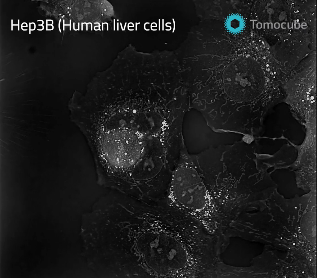
Holotomography (HT) is the most versatile label-free live cell analysis platform providing unprecedented precision of three-dimensional subcellular information.
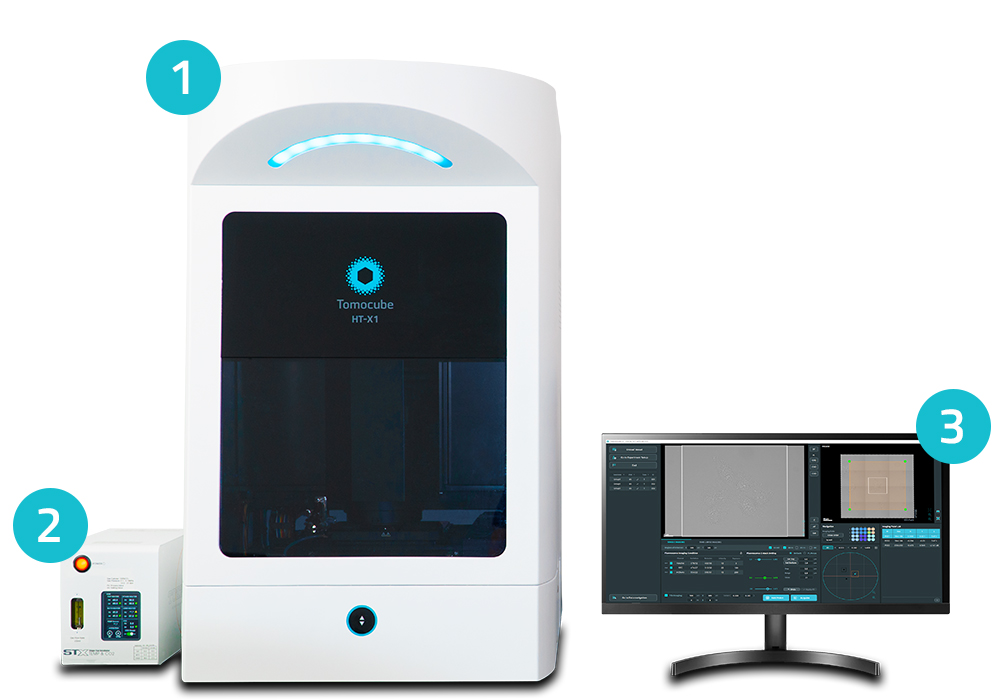
The now on-demand webinar introduces Tomocube’s HT-X1, the first ever holotomography technique to use a low-coherent light source with multiple beam patterns to obtain quantitative 3D Refractive Index (RI) information thereby minimizing interference noise, and eliminating the need for a calibration step for image acquisition.
The motorized stage allows for tiling and multi-point analysis within each well, in addition to moving between wells. A built-in incubation system completes the live-cell imaging setup. In addition to quantitative RI information, users can correlate holotomography images with 4 channel of correlative 3D fluorescence. The applications are similar to other holotomography imaging systems, although the HT-X1 is an especially powerful tool for:
Confluent & sensitive live cells: primary cells including stem cell and neuronal cells,
Monitoring of multiple samples
Observing thicker samples
Nanomaterial delivery: better resolution without speckle noise
We at Nexus would like to wish everyone a happy new year, and announce that we will be attending the Advanced Imaging Methods Conference at UC Berkeley this January 24th – 26th. If you haven’t registered yet, the opportunity is still available here. We are excited to showcase Tomocube’s new automated, label-free imaging system, the HT-X1.
For those of you who would like to learn more about the HT-X1, we will be hosting a pre-conference webinar to highlight the event and the system this Friday the 13th at 11am PST. Register here – even if you can’t make the conference it will still be a great opportunity to learn more! The MIC at Berkeley has also given us a dedicated space to house the system for the week surrounding the conference. Live demonstrations will be held for any interested users from January 18th – February 3rd. Don’t miss your chance to test your samples and book a personal visit. See below!
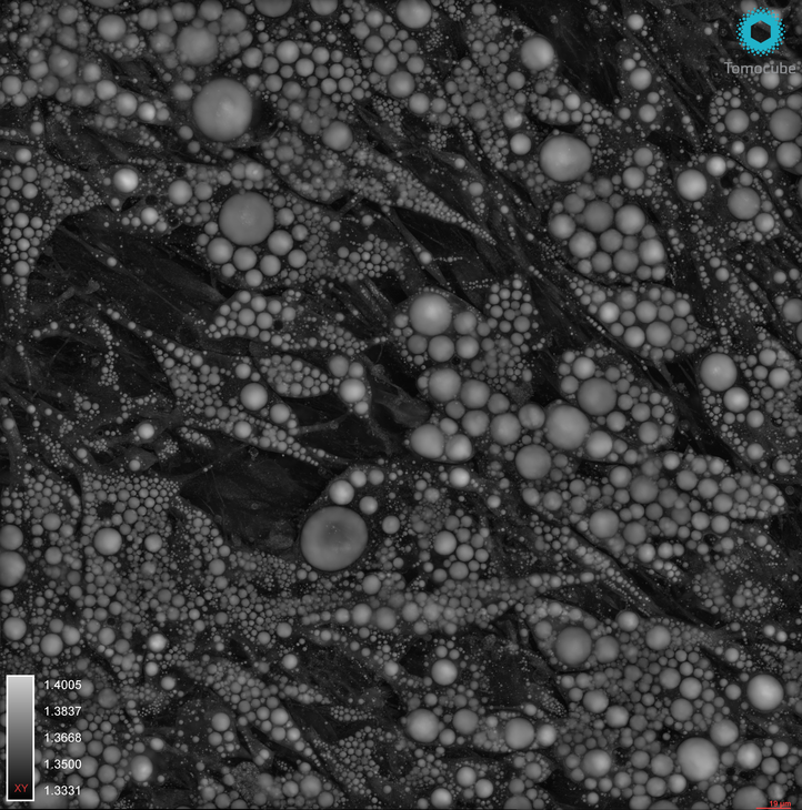
Maximum-Intensity Projection Video of a Label-Free Holotomographic Image
The HT-X1 is the latest of Tomocube’s label-free microscopes. The system adapts the ODT technique to achieve compatibility with standard multi-well plates, while still retaining the many advantages of label-free holotomography. With a large capture field, multi-point and stitching capabilities, the HT-X1 scales the amount of data acquisition by multiple orders of magnitude. 3D fluorescence capabilities and a fully integrated incubation system provide true multi-dimensional imaging across time, space and modality
PHI’s (Phase Holographic Imaging) non-invasive HoloMonitor® M4 lets you continuously image, monitor and, analyze single cells as well as cell populations directly inside the incubator without any labels or stains. It is a compact microscope that allows continuous digital holography imaging inside an incubator for tracking cells in real-time.
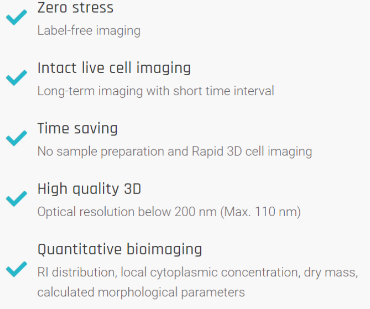
The Holomonitor® App Suite Software is an integrated solution with cell biological different application modules including:
Guided End-point Assays to quickly assess cell count and cell culture quality
Cell counting
Cell quality control
Guided Kinetic Assays for kinetic results that are publication-ready
Kinetic Cell Proliferation
Kinetic Cell Motility
Kinetic Dose Response
Wound Healing
In-dept Analysis Assays for single cell tracking for the analysis of individual cell and cell populations
Single cell tracking
Cell morphology
Automatic single cell tracking saves time
The Holomonitor® AppSuite software tracks all cells with great precision, including cell division detection and assigning daughter cells to their cell family. This gives you insights into the entire family tree of a cell without any cellular markers.
The digital holography enables automatic collection of data on an individual cell within the image, giving insights in tracking, cell behavioral, or morphological changes.
Analyze both cell movement and morphological changes
With Holomonitor®,you can follow cells over time inside the incubator. With a single sample, multiple behavioral and morphological changes in the cell culture can be studied. In a single experiment, you can examine cell movement plots over time as well as study the changes in cell morphology features within a single celland/ora cell population.
Improved data export allows deeper insights
With the App Suite software, you can export the single cell tracking analysis into a spreadsheet for further analysis. This data allows the study of morphological features, as well as cell cycle length etc.
Benefits of HoloMonitor® for Live Cell Imaging
Cut down on analysis time, costs and valuable cells
Expensive consumables are not needed
Operation is label-free
No phototoxicity
Cells can be used for other experiments after imaging
Export data to a spreadsheet with a single click
Faster tracking adjustments
Optimized non-biased results
Automated analysis keeps user bias to a minimum
Continuous cell monitoring
Real-time results
Direct, quantitative measurements
Whether you need live cell imaging for single cells or cell populations, read more about the PHI Holomonitor® at Nexus Scientific or call (857) 217-0936.The PHI Holomonitor® allows continuous imaging, monitoring and quantitative analysis of both single cells and populations of cells directly inside the incubator without any labels or stains.
Sports have been played for ages. However, competitive sports require enhanced athletic performance to improve the odds of winning. Athletes and coaches are always looking to improve sports performance. Enhancing performance requires a thorough understanding of many internal or external factors.
Most athletes are aware of internal factors, like genetics and muscle fiber type, and external factors, like the environment. But research shows us there is more to performance. One such factor is ‘Oxidative Stress’.
Why is oxidative stress harmful?
Oxidative stress refers to the difference of free radicals and antioxidants in the body. Free radicals are molecules that easily react with other molecules, causing oxidation reactions. These are usually balanced by antioxidants in the body. But when there’s the free radical activity far exceeds the antioxidant activity, you experience oxidative stress. As a result, the free radicals start affecting lipids, DNA, and proteins, causing premature aging or diseases, like heart disease, diabetes, inflammation and more.
Sports and exercise increase the production of free radicals. Exercise leads to an increased demand for oxygen, especially in the muscles, altering blood flow to the various organs. Exercise also causes muscle injury, leading to entry of phagocytes at the site. All these physiological changes add to the production of free radicals, causing oxidative damage to biomolecules.
An interesting fact to know is a moderate, short-term increase in free radicals is good for the body as it aids physical adaptation. This is possible with a good physical activity program. But excessive oxidative stress could impact an athlete’s performance in several ways.
Muscle damage
Muscle injuries are common while training. But did you know that damaged muscle fibers cause infiltration of phagocytes that break down the damaged tissue and further attract more white blood cells to the site.
This can ideally cause regeneration of muscle fibers. However, in case of excessive activity, it may lead to chronic inflammation, improper healing, and, in some cases, scar-tissue formation.
Motor skills
Retired professional athletes, especially those who played contact sports, have been seen to be at a higher risk of neurological diseases, like Alzheimer’s and ALS.
Atherosclerosis
Cholesterol is required in the production of several biomolecules, such as hormones, and coenzymes. It becomes harmful when free radicals oxidize it. In that case, it gets embedded in walls of the blood vessels, preventing normal blood flow. A combination of this and oxidative stress can lead to atherosclerosis.
Can Oxidative stress be prevented?
Oxidative stress is necessary to some extent, so it can’t be completely eliminated from sports. As an athlete, the next best thing is oxidative stress tracking for recovery throughout a training cycle to make sure you are creating positive adaptations without causing cell damage.
Fortunately, the O2Score device, available at Nexus Scientific, makes it possible to scientifically quantify the body’s response to training. Athletes and coaches can make sure the training loads and recovery methods are optimal, and performance can be maximized, without causing excessive oxidative stress and the risk of injury or fatigue.
Thank You
Your form has been successfully submitted.
Get A Quote
Kataoka Cell Processor
PHI Downloadable Website Brochures
Our Spring Sale Has Started
You can see how this popup was set up in our step-by-step guide: https://wppopupmaker.com/guides/auto-opening-announcement-popups/