Biopixlar
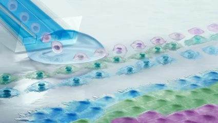
Fluicell’s Biopixlar is a 3D single-cell bioprinting platform with its unique high resolution and precise capability of positioning cells directly on cell culture media without the need for any bioink. This allows the researchers to build biological tissues for drug development, disease understanding, and regenerative medicine research.
Some of the benefits of using Biopixlar bioprinter includes:
- Single cell resolution
- High precision and reproducibility
- >95 % cell viability
- Printing up to 3 different cell types from the same print-head
- Real-time monitoring with the fully integrated multi-color fluorescence imaging setup
- Ideal for valuable cell sources such as stem cells, primary cells and patient biopsies
Biopixlar Platform
Fluicell’s Biopixlar platform consists of a bioprinter, a software, a gamepad, all situated on an optical table.
The bioprinter is equipped with micromanipulator arm, a motorized stage, a holder that connects the microfluidic printhead with a precision pressure controller, and Fluicell’s microfluidic printhead that is made from a medical-grade elastomer and includes wells for containing cell suspension, reactant solutions and for collecting waste. Furthermore, multi-color fluorescence imaging setup allows real-time monitoring of the printing process and post-print analysis. The system also has built-in environmental air-flow control, printhead heating, illumination of both the full chamber and the surrounding of the printing area, and optional UV light sterilization.
With Biopixlar’s software, the user can control the positioning of the printhead, cell type, deposition mode, and printing rate as well as adjusting the environmental controls. The gamepad allows the user to position the printhead, and deposit cells with the press of a button. A graphical user interface is also included for design of simple 2D structures.
Biopixlar Tissue Models
Biopixlar is perfect for:
- Producing human-like tissue replicas
- Understanding of specific diseases
- Testing of drug compounds
- Translating new therapies and technologies to patient
Biopixlar Printing Examples
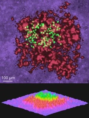
3D Tissue Model
Top image: A fluorescence overlay of the 3D tissue model printed, with skin cancer cells as a base line (A431, in purple), epithelial cells as a middle layer (HaCaT, in red) and skin cancer cells as a top layer (A431, in green). Bottom image: an alternate representation of this layered visualization.
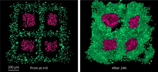
Skin Cancer Model
Fluorescence microscopy images of four printed patches of skin cancer cells (A431, in pink) surrounded by epithelial cells (HaCaT, in green) taken at t=0 and 24h after printing.
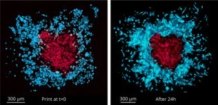
Liver Model
Fluorescence microscopy images of a printed patch of liver cancer cells (HepG2, in red) surrounded by fibroblasts (3T3-J2, in blue) taken at t=0 and 24 hours after printing.
Biopixlar Applications
Here are some Biopixlar applications